Fig. 2.
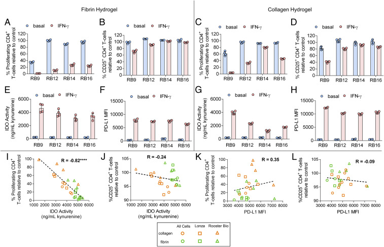
Immunosuppressive capacity of IFN-γ–licensed MSCs on hydrogel biomaterials. (A) The percent of proliferating and (B) percent of CD25+ CD4+ T cells in the presence of MSC lines, with and without IFN-γ licensing, cultured on fibrin. (C) The percent of proliferating and (D) percent of CD25+ CD4+ T cells in the presence of MSC lines, with and without IFN-γ licensing, cultured on collagen. (E) Activity of IDO, as measured by concentration of kynurenine in conditioned media, and (F) mean fluorescence intensity (MFI) of PD-L1 of various MSC cell lines, with and without IFN-γ licensing, cultured on fibrin. (G) Activity of IDO and (H) MFI of PD-L1 of various MSC cell lines, with and without IFN-γ licensing, cultured on collagen. (I and J) Correlation between IDO activity with (I) proliferating CD4+ T cells and (J) CD25+ CD4+ T cells of IFN-γ–licensed MSC cell lines cultured on fibrin and collagen. (K and L) Correlation between PD-L1 MFI with (K) proliferating CD4+ T-cells and (L) CD25+ CD4+ T cells of IFN-γ–licensed MSC cell lines cultured on fibrin and collagen. Data are presented as means ± SD. Significance is denoted by ****P ≤ 0.0001 by two-tailed Spearman’s rank correlation.

