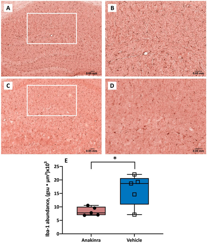
Figure 5.
Expression of Iba-1 immunoreactivity in the CA1 region of hippocampus in mice with autoimmune seizures. (A-D) Representative Iba-1 immunostaining images of the CA1 region of vehicle-treated (upper panel) and anakinra-treated (lower panel) mice with seizures induced by anti-NMDAR antibodies at 10 X (A, C) and 20 X (B, D). (E) Anakinra reduced the expression of Iba-1 in the CA1 region of hippocampus in mice with seizures. The abundance of Iba-1 labeling in the CA1 region was determined as the sum of the products of mean pixel intensity (gsu) and area of each event (μ2) in a fixed scan area. N=5 (anakinra-treated), n=5 (vehicle-treated). * p<0.05, Student’s t-test. Error bars represent mean ± SEM.

