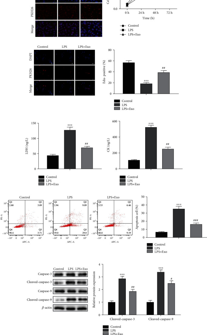
Figure 2.

Effects of MSCs-Exo on the proliferation and apoptosis of cardiomyocytes in vitro. (a) Red dye PKH26-labeled MSCs-Exo was detected in LPS-induced cardiomyocytes H9C2 cells. (b) The cell viability was detected by CCK-8 assay. (c) The cell proliferation was examined by EdU assay. (d) The release levels of LDH and CK were measured by LDH detection kit and CK detection kit, respectively. (e) The cell apoptosis was assessed by flow cytometry assay. (f) The protein levels of caspase-3, cleaved-caspase-3, caspase-9, and cleaved-caspase-9 were detected by Western blot assay. ∗∗∗P < 0.001, #P < 0.05, ##P < 0.01, ###P < 0.001 vs. the control group.
