Fig. 1.

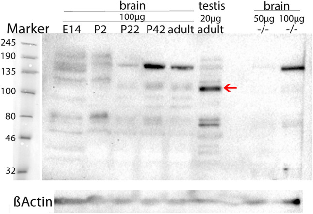
Western blot imaging of MORC1 protein [anti-MORC1 from rabbit (1:500; 14,080–1-AP, Proteintech)] in rat brain at the age of E14 (pooled from 5 embryos), P2 (pooled from 4 pups), P22, P42, adult (P > 60), and protein from an adult rat testis (1 animal each). Moreover, MORC1 protein was stained in one adult female C57BL/6 N Morc1 (−/−) brain using 50 µg and 100 µg protein. Imaging was performed with the ChemiDoc™ MP Imaging System (BIO-RAD). A prominent band was detected at 110 kDa (red arrow) in all rat samples but not the Morc1 (−/−) sample. Beta-actin staining revealed prominent bands at 45 kDa (ßActin) in all samples
