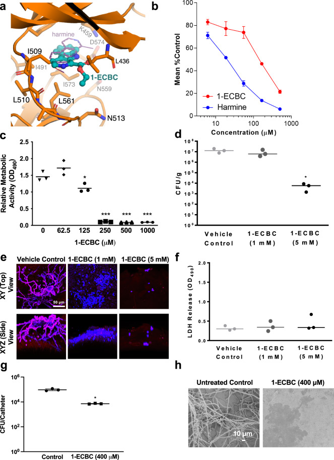
Fig. 6. 1-ECBC inhibits virulence of C. albicans.
a Homology model of 1-ECBC bound to the Yak1 kinase domain. Critical residues important for the interaction are depicted. For comparison, harmine bound to mammalian DYRK1A (PDB 3ANR70) is shown as a thin purple line. b 1-ECBC and harmine inhibit Yak1 activity in a concentration-dependent manner. Error bars represent ±SEM of five technical replicates. Data are representative of two biological replicates. c 1-ECBC prevents the development of C. albicans biofilms in vitro. Biofilms were grown for 24 h in the absence or presence of 1-ECBC as indicated. The XTT dye was used to evaluate metabolic activity. Data is an average of five technical replicates and representative of two biological replicates. Measure of center represents the mean of the data. Significance of differences between wild type and all treatment groups was determined by one-way ANOVA; *p value < 0.0136, ***p value < 0.0001. d 1-ECBC reduces tissue fungal burden on a murine vaginal explant (n = 3). Significance of differences between the vehicle control and all compound treatment groups was determined by one-way ANOVA; *p value = 0.0284. Measure of center represents the mean of the data. e Fluorescence microscopy of C. albicans biofilms on vaginal explants treated with 1-ECBC. Blue indicates calcofluor white staining and red indicates concanavalin red staining. All images at ×600 magnification (scale bar is 50 μm). Data represent two biological replicates. f 1-ECBC prevents biofilm formation without causing tissue damage in vaginal explants (n = 3). LDH release of vaginal tissue following inoculation with wild-type C. albicans and treatment with or without 1-ECBC for 24 h. Measure of center represents the mean of the data. g 1-ECBC (400 μM) prevents C. albicans biofilm formation in a rat catheter model35. Serial dilutions of catheter fluid were plated for viable fungal colony counts in triplicate. Data are presented as mean colony forming units (CFU) per catheter for three technical replicates: one female rat catheter per treatment (*P = 0.015, paired two-sided t test). Measure of center represents the mean of the data. h Scanning electron microscopy (SEM) images of biofilms formed on catheters. Images are representative of catheters from three biological replicates. All source data for this figure are provided as a Source Data file.

