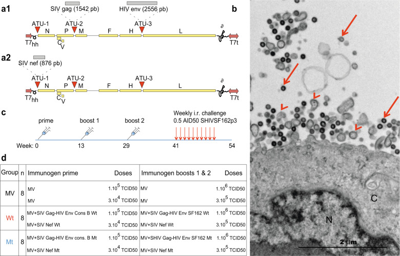
Fig. 1. Vectors, VLP electron microscopy, and vaccine regimen.
a1 pMVSchw vector with ATU2 containing SIV gag gene and ATU3 containing HIV env gene WT or mutated. HIV env genes are HIV env cons B subtype for the prime vector and SF162 subtype for boosts 1 and 2. a2 pMVSchw vector with ATU1 containing SIV nef gene WT or mutated. HIV env and SIV nef genes were mutated at their immunosuppressive domains. The MV genes are indicated as follows: N (nucleoprotein), P (phosphoprotein), V and C proteins, M (matrix), F (fusion), H (hemagglutinin), L (polymerase), T7 (T7-RNA polymerase promoter), T7t (T7-RNA polymerase terminator), δ (hepatitis delta virus ribozyme). b Electron microscopy image of Vero cells infected by recombinant MV-SHIV Wt virus (MOI of 0.01, MV-SIV Gag-HIV Env). N nucleus, C cytosol. Arrowheads indicate MV viral particles and arrows gag-forming VLPs. c, d Summary of vaccine: immunization schedule, and repeated low dose of intrarectal SHIVSF162P3 challenges. Prime and boost 1 immunizations were subcutaneous and boost 2 was both subcutaneous and intranasal. Subcutaneous inoculations were performed at two distant sites in the back of animals.

