Figure 5.
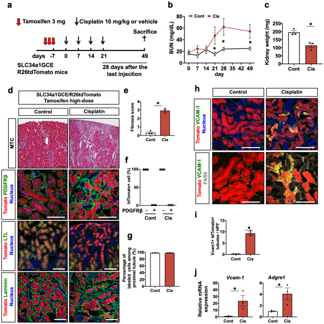
Maladaptive tubular repair and interstitial fibrosis after repeated administration of 10 mg/kg of cisplatin. (a) Experimental scheme. Repeated administration of 10 mg/kg of cisplatin to the SLC34a1GCE R26tdTomato mice after labeling of proximal tubular epithelial cells by multiple injections of high-dose tamoxifen. The kidneys were assessed at a later phase, 28 days after the last injection. (b) Chages in BUN level during and after repeated administrations of cisplatin. (c) The average kidney weight in the cisplatin and control groups. (d) Histological analysis of the kidneys in the cisplatin and control groups. Masson’s trichrome and immunostaining of PDGFRβ, LTL, and laminin. (e) Quantification of the fibrosis score. (f) Quantification of PDGFRβ positivity in the tdTomato + cells. tdTomato + tubules never merged with PDGFRβ. (g) Quantification of labeled cells. Labeled cells were not diluted in the cisplatin model. (h) Immunostaining of vcam-1 and F4/80. (i) Quantification of vcam-1 + tdTomato + tubules. (j) Quantitative PCR of whole kidneys for vcam-1 and adgre1 genes. Data are the average ± SE. *P < 0.05. Scale bar = 100 μm in MTC and 50 μm in others.

