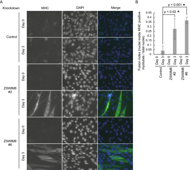
Figure 2.
Accelerated myotube formation with ZSWIM8 knockdown. (A) Growing (day 0) or differentiated (day 3) control or ZSWIM8-knockdown (#2 or #6) C2C12 cells were immunostained with an anti-myosin heavy chain (MHC) antibody. Nuclei were stained with DAPI. Scale bar, 20 μm. MHC-positive and multinucleated cells were considered myotubes. (B) Quantification of fusion indexes of (A). The number of nuclei inside MHC-positive myotubes was divided by total nuclei in different fields. Data represent the mean ± SD; n = 150. Asterisk indicates statistical significance compared to the control sample.

