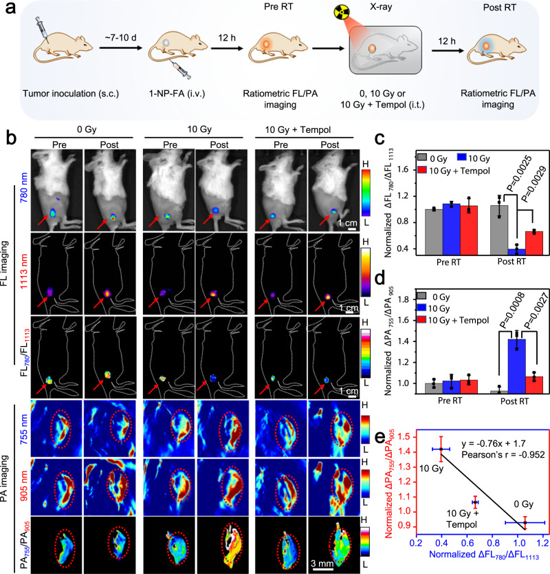
Fig. 6. Ratiometric bimodal imaging of •OH production in 4T1 tumors undergoing X-ray RT.
a Schematic for noninvasive ratiometric fluorescence/photoacoustic (FL/PA) imaging of •OH in 4T1 tumor-bearing mice upon radiotherapy (RT) with X-ray. 4T1 tumor-bearing mice were intravenous (i.v.) injected with 1-NP-FA (1.68/0.05/0.6 mM 1-Br-Et/NIR775/IR1048, 200 μL). After 12 h, the FL and PA images were acquired (Pre RT). The tumors were then unirradiated (0 Gy) or irradiated with X-ray (10 Gy, 1.0 Gy min−1 for 10 min); to inhibit tumor •OH, the mice were intratumorly (i.t.) injected with tempol (50 mg kg−1), and then irradiated with X-ray (10 Gy). After another 12 h, the FL and PA images were acquired (Post RT). b FL, PA, and corresponding ratiometric FL or PA images of 4T1 tumors following indicated treatment. Red arrows and circles indicate the tumor locations. c Normalized ΔFL780/ΔFL1113 ratios and d normalized ΔPA755/ΔPA905 ratios in 4T1 tumors following indicated treatment. e Plot of the normalized ΔFL780/ΔFL1113 ratios versus normalized ΔPA755/ΔPA905 ratios shows a good correlation (r = −0.952) between them in 4T1 tumors upon indicated treatment. Data are presented as mean ± s.d. (n = 3 independent mice). Statistical differences were analyzed by Student’s two-sided t-test. Source data are provided as a Source Data file.

