
Figure 6. RPE regeneration in zebrafish initiates in the periphery and proceeds inward.
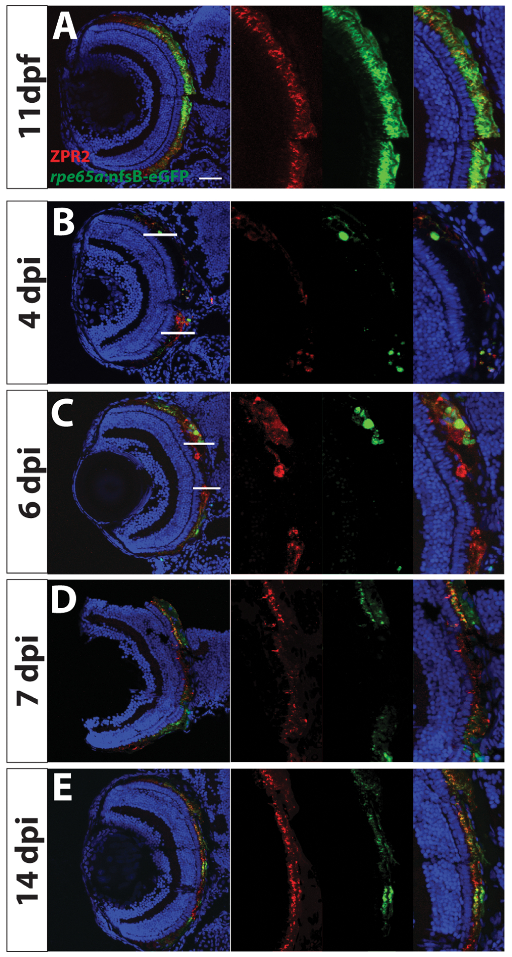
Transverse sections of unablated larvae stained for the RPE marker ZPR2 (A) at 11dpf. Ablated eyes stained for ZPR2 at 4, 6, 7 and 14dpi (B-E). Green=eGFP, blue=nuclei, red=ZPR2. eGFP+ RPE appears in the periphery at 4dpi (marked by lines at dorsal and ventral margins in B,C). As regeneration proceeds, eGFP+ RPE extends further toward the eye center, and the leading tip of the regenerated monolayer often consists of both immature and mature RPE (ZPR2+/eGFP− cells in C). By 7dpi, ZPR2+ RPE is present throughout the RPE (D). By 14dpi, mature eGFP+/ZPR2+ RPE cells are present throughout the RPE (E). Dorsal is up and distal is left. Abbreviations: RPE, retinal pigment epithelium; dpf, days post-fertilization; dpi, days post-injury. Scale bar = 40mm. Figure modified from Hanovice et al., 2019.