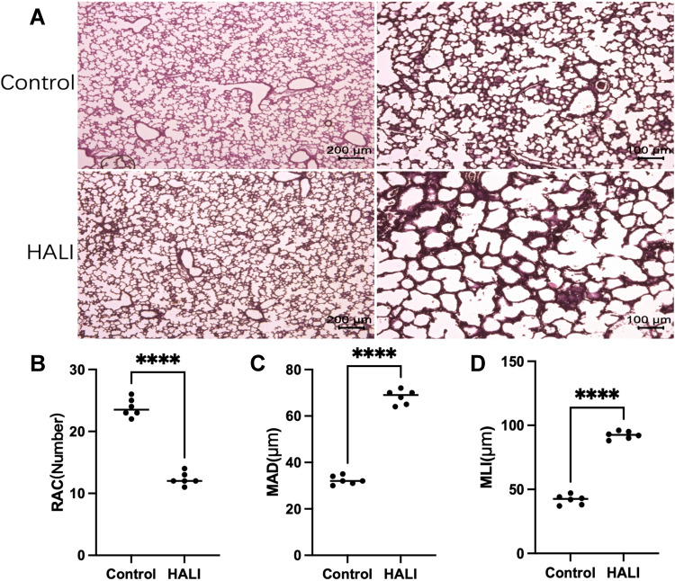
Figure 1.
(A) In HE staining, the lung tissue structure in the HALI group showed a decrease in the number of alveoli, an increase in the volume of the alveolar cavity, and a thickening of the alveolar septa compared with the control. Scale bars = 200 μm, 100 μm. (B) Semi-quantitative pathology score of RAC in lung tissues. (C) Semi-quantitative pathology score of MAD in lung tissues. (D) Semi-quantitative pathology score of MLI in lung tissues. The values are the mean ± SD; n = 6 nonoverlapping fields/group; ****P < 0.0001.

