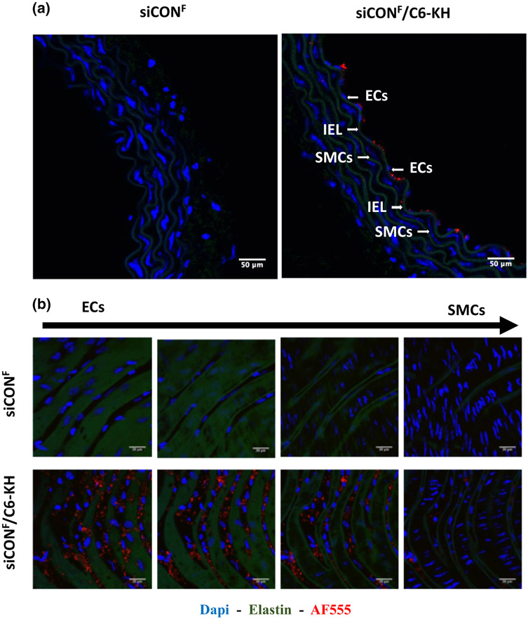
FIGURE 3.
siCONF delivery to endothelial layer. Abdominal aortas of C57Bl6 mice were dissected out and incubated ex vivo with siCONF-C6-KH or naked siCONF for 1 h, followed by either frozen cross-section (a) or en face (b) imaging by confocal microscopy. Blue DAPI for nuclear, Green auto-fluorescence for elastic laminas, Red AF555 for siCONF are shown.

