Figure 7.
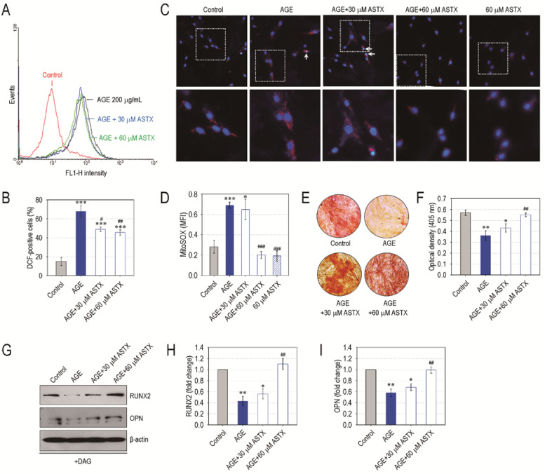
ASTX inhibits oxidative stress and increases mineralization in AGE-treated hPDLCs. The hPDLCs were incubated in growth medium supplemented with and without AGE (200 μg/mL), ASTX (30 or 60 μM), or both for 48 h. (A) ROS accumulation in the cells was determined by flow cytometry using DCFH-DA, and (B) the DCF-positive cells (%) were calculated (n = 5). (C) The level of mitochondrial superoxide in the cells was determined by staining them with MitoSOX Red and DAPI, and (D) the MFI specific to the red dye was calculated using ImageJ software (n = 5). Alternatively, the hPDLCs were incubated in DAG-supplemented osteogenic medium in the presence and absence of AGE, ASTX or both. (E) After 14 days of incubation, the cells were stained with Alizarin Red S, and (F) the red dye-specific optical density in the cells was determined using a microplate reader (n = 5). (G) The protein levels of osteogenic markers in the cells exposed to AGE, ASTX, or both were analyzed by immunoblot assay 5 days after incubation, and the band intensities of (H) RUNX2 and (I) OPN were calculated (n = 4). * p < 0.05, ** p < 0.01, and *** p < 0.001 vs. the untreated control hPDLCs. # p < 0.05, ## p < 0.01, and ### p < 0.001 vs. the hPDLCs exposed to AGE alone.

