Figure 8.
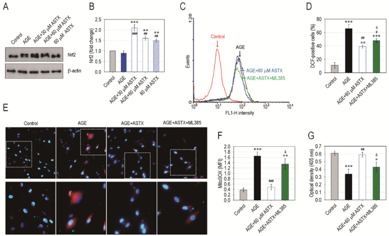
ASTX inhibits oxidative stress and increases proliferation in AGE-exposed hPDLCs by upregulating Nrf2 pathway. The hPDLCs were incubated in growth medium supplemented with and without AGE (200 μg/mL), ASTX (30 or 60 μM), or both. (A) After 48 h of incubation, the protein level of Nrf2 in the cells was determined by immunoblot assay, and (B) the Nrf2-specific band intensity (fold change to control) was calculated after normalizing its level to that of β-actin (n = 4). The hPDLCs were also exposed to AGE, ASTX, or both with and without the pretreatment with 5 μM ML385. After 48 h of incubation, (C) the DCF-specific signal and (D) the DCF-positive cells (%) in the hPDLCs were calculated by flow cytometry (n = 5). After the same time, (E) immunofluorescence assay was also performed using MitoSOX Red and DAPI, and (F) the red dye-specific MFI among the experiments was compared after calculating the levels using ImageJ software (n = 5). (G) In addition, proliferation rate of the cells was determined at the end of 48 h incubation using a CCK-8 assay kit. * p < 0.05, ** p < 0.01, and *** p < 0.001 vs. the untreated control hPDLCs. # p < 0.05, ## p < 0.01, and ### p < 0.001 vs. the hPDLCs exposed to AGE alone. & p < 0.05 vs. the cells exposed to ASTX in combination with AGE.

