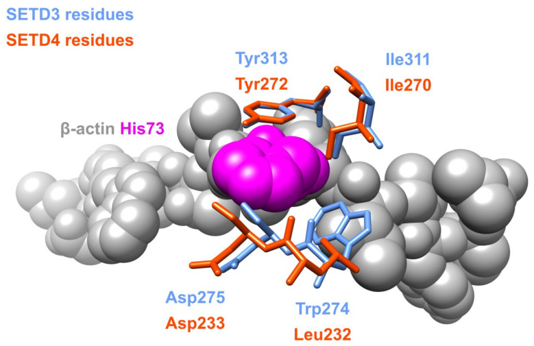
Figure 4.
Structural alignment of SETD3 amino acid residues interacting with H73 of β-actin and conserved residues of SETD4. The image was created in UCSF Chimera 1.15 software utilizing the coordinates deposited in Protein Data Bank file 6ICV and the SETD4 structure predicted by AlphaFold [41] using UniProt Q9NVD3 record as an input. Structural alignment was calculated using the MatchMaker tool in UCSF Chimera 1.15 software [36].

