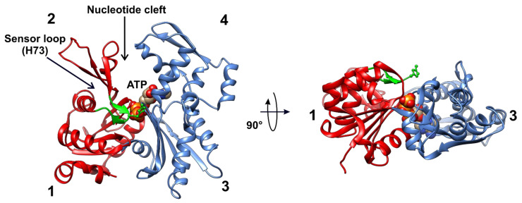
Figure 5.
Structures of human β-actin. Ribbon representations of the structures of the actin monomer are shown in different projections. The actin molecule consists of small and large domains (red and blue, respectively), and each one is divided further into two subdomains: 1, 2, and 3, 4, respectively. ATP (or ADP) binds to the cleft between subdomains 2 and 4. The methyl-accepting H73 is located in a sensor loop spanning P70 to N78 (green). This residue is exposed to the surface of the actin monomer and seems to be easily accessible for SETD3. The model was prepared using UCSF Chimera [36] from the Protein Data Bank structures of β-actin (2BTF).

