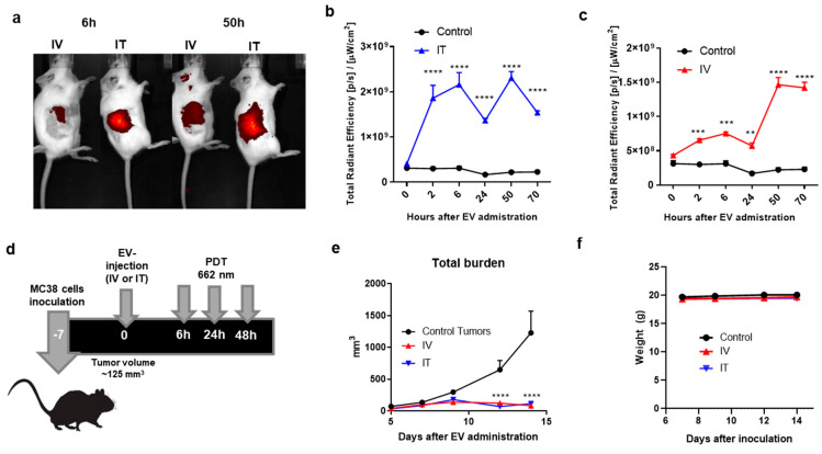
Figure 4.
Biodistribution and effect of EV-ZnPc after PDT in vivo. Mice were inoculated subcutaneously with 0.5 × 10⁶ MC38 cells in 200 μL PBS in the right flank. At day 7, when the tumors were an average size of ~125 mm3, EV-ZnPc were injected intravenously (IV) or intratumorally (IT). Non-invasive fluorescence imaging was performed at 2, 6, 24, 50, and 70 h after injection using the IVIS fluorescence spectrometer. (a) Representative NIR700 fluorescence imaging in the tumor area of mice at 6 and 50 h after injection. Fluorescence intensity of the NIR800 signal after (b) IT or (c) IV injection. (d) Schematic representation of protocol used for PDT assay: mice were inoculated with MC38 cells in the flank and injected with EV-ZnPc as mentioned above. PDT was performed by illumination of the tumor on the right flank with 662 nm light at 116 mW/cm2 for 116 J/cm2 over 1000 s. (e) Tumor growth curves and (f) weight of the animals were evaluated over 14 days after inoculation. Data are means ± SEM from n = 4 mice per condition. **** p < 0.0001; *** p < 0.001; ** p < 0.01.

