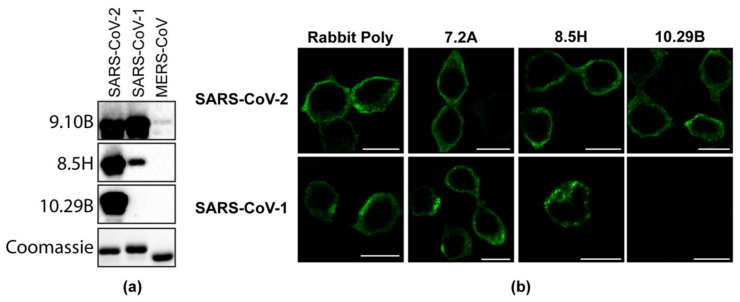
Figure 2.
(a) Western blot showing purified N proteins from SARS-CoV, SARS-CoV-2, and MERS-CoV probed with monoclonal N antibodies 9.10B, 8.5H, and 10.29B. A Coomassie-stained polyacrylamide gel shows the equal loading of the proteins. (b) 293T cells transfected with a plasmid expressing SARS-CoV-2 or the SARS-CoV N protein were fixed at 24 h post-transfection and probed with the rabbit polyclonal serum, or 7.2A, 8.5H, and 10.29B mouse monoclonal antibodies.

