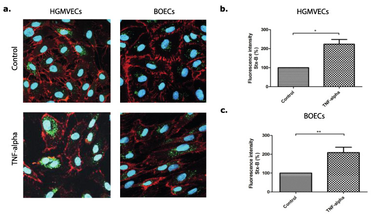
Figure 2.
The expression and binding of Alexa 488 labelled Shiga toxin subunit B (Stx-B) on the cell surface of primary HGMVECs and BOECs. (a) Immunofluorescent images of Stx-B on HGMVECs and BOECs pre-incubated without (control) or with 10 ng/mL of TNFα for 24 h. Stx-B in green and CD-31 in red. The nucleus is stained in blue; 400× Magnification by confocal microscopy. Flow cytometry of the binding of Stx-B on the cell surface of (b) primary HGMVECs or (c) BOECs pre-incubated without (control) or with 10 ng/mL of TNFα for 24 h. Pre-incubation of TNFα resulted in an increase of Stx-B binding on the endothelial cell surface of both HGMVECs and BOECs. Statistically significant differences are indicated with single (p < 0.05) or double (p < 0.01) characters, respectively.

