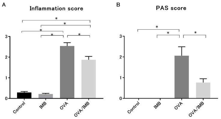
Figure 8.
Histological examination of lung tissues. Lung inflammation was evaluated by HE staining, and hyperplasia of goblet cells was assessed by PAS staining. The semi-quantitative scores of inflammatory infiltration and goblet cell hyperplasia were significantly lower in the OVA/IMB group than in the OVA group (A,B). Values represent the mean ± standard error of the mean. * p < 0.05.

