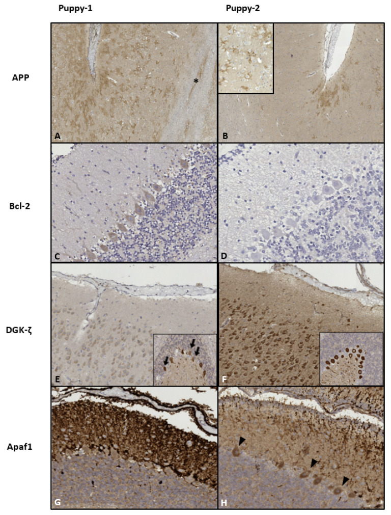
Figure 1.
Immunohistochemicalpatterns of the implemented protein markers. (A,B): Neocortex. β-amyloid precursor protein/APP localized to neuronal synaptic membranes, multifocally enhanced against a light background in the superficial cortical layers. Intense staining was diffuse in Puppy-1, with affected white matteraxons (*). In Puppy-2, strong intensity was limited to the sulci; magnification ×50. Inset in (B) (×200): detail of arborized synaptic pattern. (C–H): apoptotic markers. (C,D): Moderate diffuse Purkinje cell cytoplasmic B-cell lymphoma related protein 2/Bcl-2-signal (with slight emphasis on gyral crowns; not shown) in Puppy-1 vs. predominantly negative cerebellum in Puppy-2. Magnification ×400. (E,F): Neocortex: “faded” appearance in Puppy-1 vs. strong cytoplasmic diacylglycerolkinase-ζ/DGK-ζ-signal in neurons and neuropil in Puppy-2; magnification ×100. Insets depict the puppies’ cerebella, whereby Puppy-1 displays more neuronal loss (arrows). In both animals, immunoreactivity was more pronounced in the gyral crowns, as opposed to the sulci (not shown). (G): Cerebellum of Puppy-1 shows intense and diffuse, predominantly axonal apoptotic protease activating factor 1/Apaf1 immunoreactivity in the molecular layer of cerebellar folia, while in Puppy-2, intensity is moderate and the pattern is multifocal to coalescing (H). The neuronal cytoplasm is negative in Puppy-1, yet there are multiple gyral foci of moderate immunoreactivity in Puppy-2 (arrowheads in (H)); Magnification ×200.

