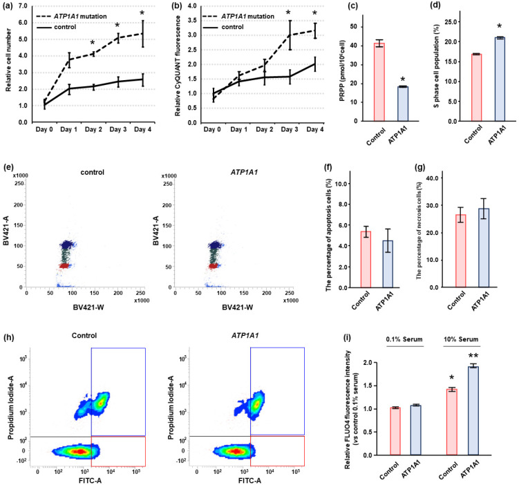
Figure 4.
Effect of ATP1A1 L104R mutation on cell proliferation in HAC15 cells. (a,b) HAC15 cells with 40–50% confluency on 96-well plates were transduced with a lentivirus ATP1A1 mutant (n = 6) or control (n = 6) vectors. The cells were cultured in DMEM/F12 containing 10% serum. The effects of the ATP1A1 mutant on cell proliferation and DNA amounts were evaluated by counting cell number using TC20 Cell Counter at the indicated times and CyQUANT Direct Cell Proliferation Assay Kit, respectively. *, p < 0.05 vs. each type of control cells. (c) After transduction of ATP1A1 mutant (n = 3) or control (n = 3) in HAC15 cells and attainment of confluency. Cells were incubated in DMEM/F12 containing 10% serum on 6-well plates for 24 h, and Phosphoribosyl Diphosphate (PRPP) levels in cells were measured by capillary electrophoresis time-of-flight mass spectrometry. (d,e) After transduction of ATP1A1 mutant (n = 3) or control (n = 3) in HAC15 cells and attainment of confluency. Incubation with DMEM-F12 containing 10% cosmic calf serum on 6-well plates for 48 h, cells were treated with 10 μM of cycloheximide for 24 h. The cells with supplemental solutions of Cell Cycle Assay Solution Blue were injected into the flow cytometer instrument, and the cells of G0/G1, S, or G2/M phase were shown in the figure with red, green, or blue, respectively. The number of cells of S phase were compared. *, p < 0.05 vs. control cells. (f–h) After transduction of ATP1A1 mutant (n = 3) or control (n = 3) and incubation with DMEM-F12 containing 10% cosmic calf serum on 6-well plates for 72 h, cells were harvested. Following Promokine Apoptotic/Necrotic Cells Detection kit (PromoCell, Heidelberg, Germany), the cells (2.5 × 10⁵) were incubated with Annexixn and Ethidium Homodimer for 15 m. The samples containing cells were injected into the flow cytometer instrument (BD FACSAriaIIu, BD Biosciences, Franklin Lakes, NJ, USA). The results of control and ATP1A1 mutation are depicted in A and B, respectively. The cells in red and blue squares were defined as apoptotic and necrotic, respectively. The apoptotic and necrotic cell number per total cell number were compared between control and ATP1A1 mutation cells. (i) After transduction of ATP1A1 mutant (n = 4) or control (n = 4) vectors in HAC15 cells and incubation with DMEM/F12 containing 0.1% serum for 24 h, cells were incubated with fresh media with or without 10% serum with 3 μM of Fluo4-AM on 96-well plates for 10 m. *, p < 0.05 vs. control cells with 0.1% serum. **, p < 0.05 vs. other three type of cells.

