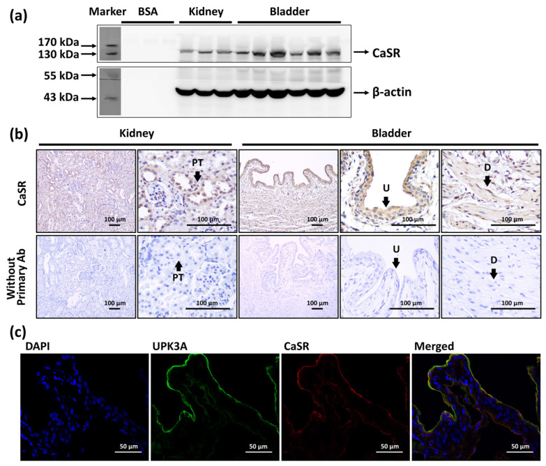
Figure 1.
The expression and location of calcium-sensing receptor (CaSR) in bladder. (a) Western blot analysis of CaSR (MW: 130 kDa) in the whole kidney and whole urinary bladder. (b) Immunohistochemistry staining of CaSR (brown signals) in the urothelium (U) and detrusor (D) of bladder, and in the proximal tubule (PT) of kidney, and the staining without applying primary antibody (Ab). (c) Immunofluorescence staining of CaSR (red), uroplakin III A (UPK3A) (green), and DAPI (blue) in urothelium of urinary bladder.

