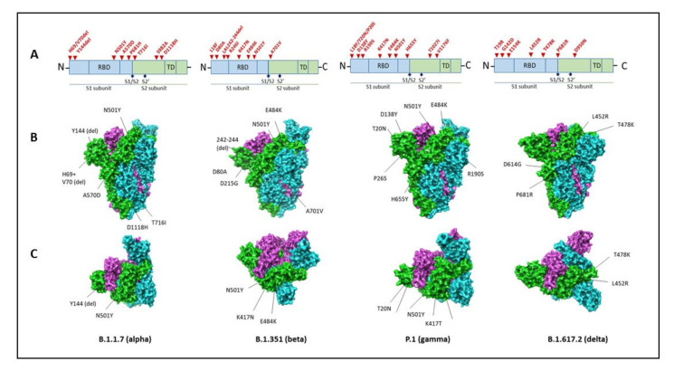
Figure 3.
Spike mutations in the variants of concern. Amino acid substitutions and their locations in the Spike protein (A). The following PDB files are used for structural illustrations: 7LWV for B.1.1.7 (Alpha); 7LYN for B.1.351 (Beta); 7LWW for B.1.1.28.1 P.1 (Gamma); and 6ZGE for B.1.617.2 (Delta). Chain A: green, Chain B: blue, Chain C: purple. (B) Side view and (C) top view. The figures were prepared using Schrodinger software, and PDB files were obtained from protein databank (https://www.rcsb.org/, accessed on 9 September 2021).

