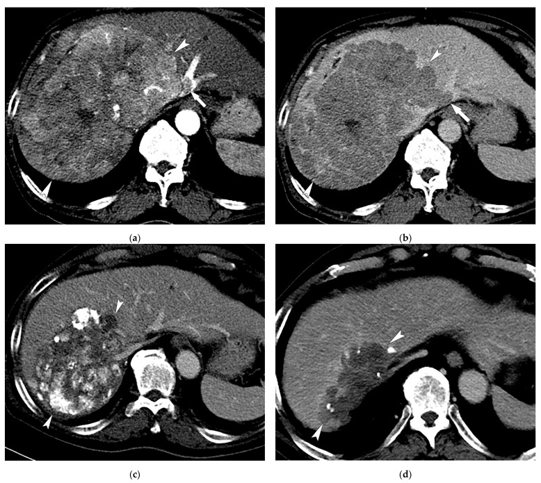
Figure 2.
Images of a 59-year-old male patient with multinodular HCCs with MVI who received TACE combined with sorafenib treatment. Contrast-enhanced axial CT images in the arterial (a) and delayed (b) phases show a huge enhancing mass (17 cm in maximal diameter; arrowheads) invading the left hepatic vein (arrows). (c) CT image at 1 year after 4 sessions of TACE combined with sorafenib therapy shows necrotic change with lipiodol uptake in the tumor and a decrease in tumor size (13 cm; arrowheads). (d) CT image at 3 years after 6 sessions of TACE shows a further decrease in tumor size without tumor viability (9 cm; arrowheads).

