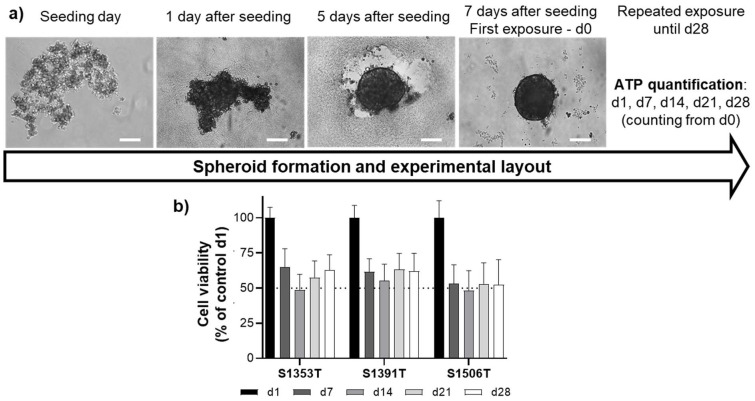
Figure 1.
(a) Timeline of spheroid generation and experimental procedure. Cryopreserved PHH were seeded in ultra-low attachment plates at a density of 1500 cells per well. The plates were centrifuged (130× g, 2 min) and kept in controlled atmosphere (37 °C, 5% CO2, humidified). The spheroids were formed 5 days after seeding and the first exposure was performed 7 days after seeding (d0). Scale bar = 100 µm. (b) Cell viability of control spheroids throughout the time-frame of the experiments. Results are expressed as mean ± standard deviation of at least 48 spheroids from each donor per time-point. ATP—adenosine triphosphate; d—day.

