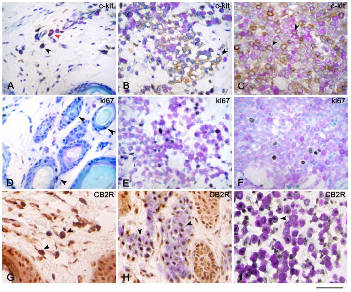
Figure 2.
Immunohistochemistry of c-kit, Ki67, and CB2 receptor with toluidine blue as the counterstain of atopic dermatitis (AD), cutaneous mastocytosis, and cutaneous mast cell tumors (MCTs). Immunohistochemistry of c-kit of AD (A), mastocytosis (B), and MCTs (C) showed cytoplasmic (black arrow) and membranous (red arrow) (A,B) and only membranous (black arrow) (C) c-kit expression of mast cells (MCs). Immunohistochemistry of Ki67 of mastocytosis (E) and MCTs (F) showed nuclear expression of MCs (E,F) visualized by toluidine blue (TB) stain, and MCs in AD were not in cycle; therefore, Ki67 stain was found only on the epithelial cells of hair follicles (black arrows) (D). Mast cells of mastocytosis (H) and MCTs (I) revealed nuclear expression of CB2 receptor (black arrows); by contrast, the MCs of AD (G) showed cytoplasmic expression of the CB2 receptor (black arrow) that hid the metachromatic reaction of TB stain. Scale bar = 50 µm.

