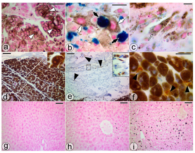
Figure 2.
Histological sections through orally treated melanoma (T4w). (a) PMC clusters (white arrowheads) with perimembranous accumulation of pigment granules. (b,c) PMCs (white arrows), NP-PMCs (thin white arrows), and large SaIONs-laden cells (black arrows) in tumor areas with nanoparticle-induced necrosis. (d) HPMCs, including some binucleate cells (square), forming overlapping layers at the periphery of the tumor (double-headed arrows). (e,f) MAC387(+) cells (black arrowheads) in juxtatumoral tissue or spread among HPMCs. (g–i) Microscopic appearance of the liver with pigment deposits in M4w, T2w, T4w groups, respectively. Perl Prussian blue (a–c), Azan Trichrome (d), and HE staining (d,g–i). Bar = 100 µm (e–i), 50 µm (a,d), 10 µm (b,c,f).

