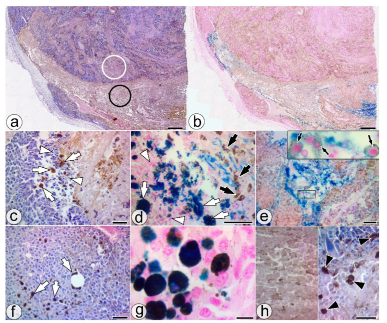
Figure 3.
Histological features of the SaIONs injected area in the ITM1w group. (a,b) Overall appearance of SaIONs-induced necrosis (black circle) in the injected area, with a different morphology from the adjacent programmed necrosis (white circle). (c,d) The border between the growing tumor and the nanoparticle-induced tumor necrosis with apoptotic bodies (white arrowheads), large MAC387- NP-PCs (white arrows), and melanin-loaded ghost cells without nuclei (black arrows). (e) SaIONs-induced tumor necrosis area with nests of viable PMCs (black arrows) surrounded by SaIONs deposits. (f,g) Melanoma tissue with large migratory NP-PCs (white arrows). (h) Higher magnification of the SaIONs-induced necrosis area (left) with pigmented ghost cells without nuclei, and the programmed necrosis area (right) with fragmented nuclei and numerous melanophages (black arrowheads). HE (a,c,f,h) and Perls’ Prussian blue staining (b,d,e,g). Bar = 10 µm (g), 20 µm (d,h), 50 µm (c,e,f), 1 mm (a,b).

