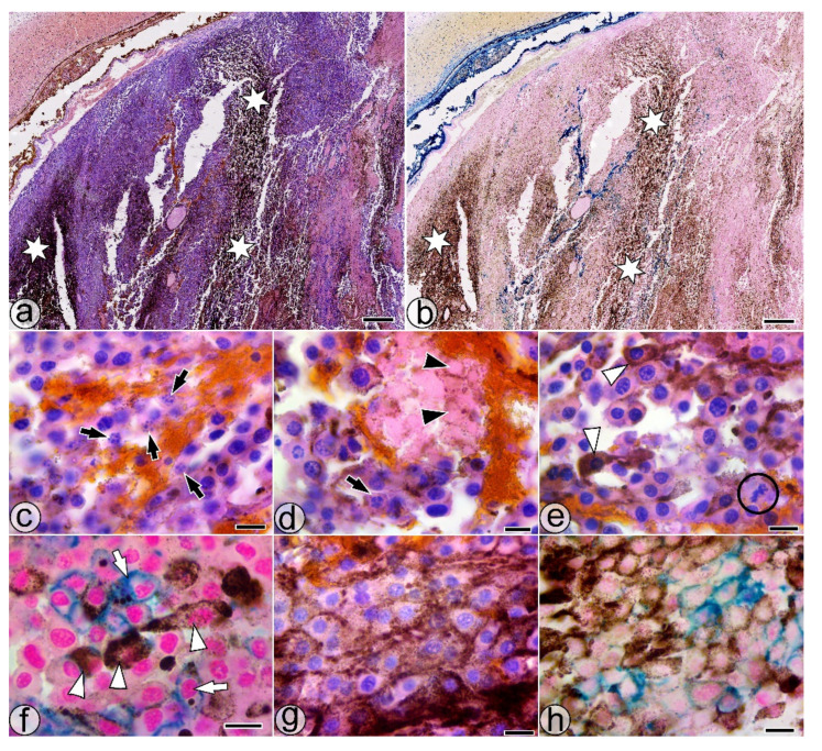
Figure 4.
Histological features of the SaIONs-injected area in the ITO1w group. (a,b) Overall appearance of the hyperpigmentation in the injected area (white stars). (c,d) Weakly pigmented melanoma cells with fragmented nuclei (black arrows) and ghost cells without nuclei (black arrowheads). (e) Area with SaIONs deposits and PMCs with varying degrees of melanin loading (white arrowheads), some in division (black circle). (f) Cluster with HPMCs (white arrowheads) and SaIONs-loaded melanoma cells (white arrows). (g,h) Deposits of pigment and SaIONs in the juxtamembranous regions of the melanoma cells. HE (a,c,d,e,g) and Perl Prussian blue (b,f,h) staining. Bar = 10 µm (c–h), 1 mm (a,b).

