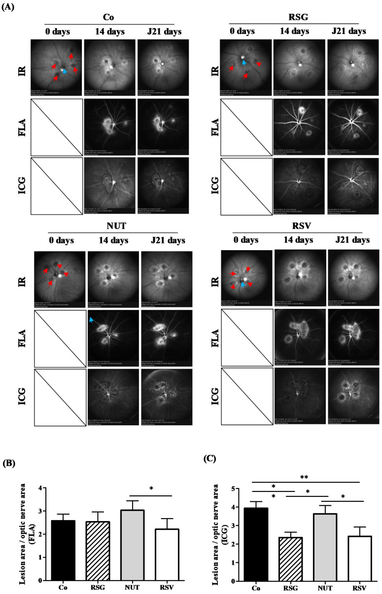
Figure 1.
RSG decreases laser-induced CNV in mice. (A) Fundus images of mice that were either untreated (Co) or supplemented with RSG (12 µM), NUT (12 µM), or RSV (20 µM) for 14 days prior to CNV induction. Infrared (IR) pictures show the four laser impacts at day 0 (red arrows) as well the optic nerve head positioned at the center of the image (blue arrows). Vascular lesions around laser spots are visible at 14 and 21 days following CNV induction on fluorescein (FLA) and indocyanin green (ICG) angiographies at inner retinal and choroidal levels, respectively. (B) Quantitative analysis of vascular lesion areas in the inner retina did not reveal significant differences between animals supplemented with RSG, NUT, and RSV when compared with untreated mice, whereas (C) a focused evaluation at the level of choroid showed significantly lower development of CNV in animals supplemented with RSG and RSV. Data are presented as means ± SEM. p values were determined by a one-way ANOVA followed by a Mann–Whitney test. * p < 0.05 and ** p < 0.01.

