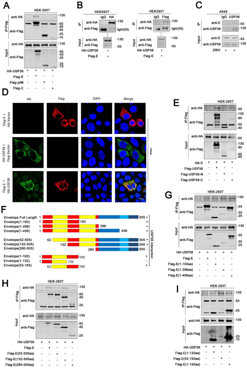
Figure 2.
USP38 binds to E protein through its C-terminal domain. (A) HEK293T cells were co-transfected with HA-USP38 and Flag-E, Flag-C, Flag-prM, cell lysates were subjected to IP using anti-flag antibody and analyzed by immunoblotting. (B) HEK293T cells were co-transfected with HA-USP38 and Flag-E. Cell lysates were subjected to IP using control IgG, anti-HA, or anti-Flag antibody. (C) A549 cells was infected with ZIKV for 48 h. Cell lysates were subjected to IP using control IgG or anti-USP38. (D) Hela cells were transfected with HA-USP38 or Flag-E, or co-transfected with HA-USP38 and Flag-E. The sub-cellular localizations of HA-USP38 (green), Flag-E (red), and nucleus marker DAPI (blue) were analyzed with confocal microscopy. Bar = 5 µm. (E) HEK293T cells were co-transfected with HA-E and Flag-USP38, Flag-N-terminal-USP38 or Flag-C-terminal-USP38. Cell lysates were subjected to IP using anti-Flag antibody and analyzed by immunoblotting. (F) Schematic diagram of the full-length E protein and truncated E proteins: E (1–193aa), E (1–296aa), E (1–406aa), E (52–505aa), E (132–505aa) E (280–505aa). (G,H) HEK293T cells were co-transfected with HA-USP38 and Flag-E, Flag-E (1–193aa), Flag-E (1–296aa), and Flag-E (1–406aa) (G) or Flag-E, Flag-E (52–505aa), Flag-E (132–505aa), and Flag-E (280–505aa) (H). Cell lysates were subjected to IP using anti-Flag antibody and analyzed by immunoblotting (G,H). (I) HEK293T cells were co-transfected with HA-USP38 and Flag-E (1–193aa), Flag-E (1–132aa), or Flag-E (53–193aa). Cell lysates were subjected to IP using anti-Flag antibody and analyzed by immunoblotting.

