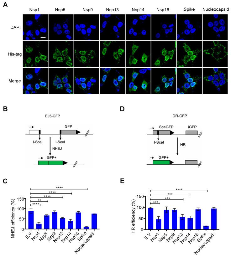
Figure 1.
Effect of severe acute respiratory syndrome coronavirus 2 (SARS–CoV–2) nuclear-localized proteins on DNA damage repair. (A) Subcellular distribution of the SARS–CoV–2 proteins. Immunofluorescence was performed at 24 h after transfection of the plasmid expressing the viral proteins into HEK293T cells. Scale bar: 10 µm. (B) Schematic of the EJ5-GFP reporter used to monitor non-homologous end joining (NHEJ). (C) Effect of empty vector (E.V) and SARS–CoV–2 proteins on NHEJ DNA repair. The values represent the mean ± standard deviation (SD) from three independent experiments (see representative FACS plots in Figure S2A). (D) Schematic of the DR-GFP reporter used to monitor homologous recombination (HR). (E) Effect of E.V and SARS–CoV–2 proteins on HR DNA repair. The values represent the mean ± SD from three independent experiments (see representative FACS plots in Figure S2B). The values represent the mean ± SD, n = 3. Statistical significance was determined using one-way analysis of variance (ANOVA) in (C,E). ** p < 0.01, *** p < 0.001, **** p < 0.0001.

