Figure 3.
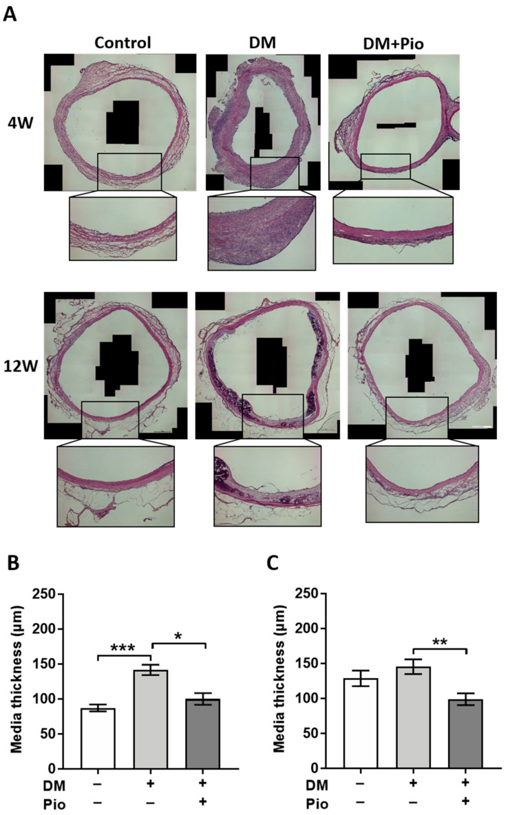
Histological results of aortic region—media thickness. For each group, tissue of n = 4 animals was stained and evaluated. (A) A representative hematoxylin eosin-stained image for each group, including higher-magnification inserts. After 4 weeks, DM had a significant thickening of the media, but it was not statistically different between the control and the pioglitazone-administered group. After 12 weeks, thickening of the media with calcification was observed in the DM group. (B) Measurement result of the media after 4 weeks. There was a significant difference in medial thickening in the DM group. (C) Even after 12 weeks, the medial thickening of the DM group was significantly different. DM, diabetes mellitus; W, weeks; Pio, pioglitazone; * p < 0.05; ** p < 0.01; *** p < 0.001. Data were analyzed using Kruskal–Wallis with Dunn’s multiple comparisons test.

