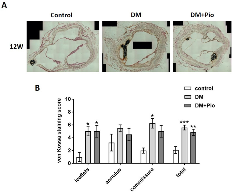
Figure 5.
Histological results of aortic region –calcification of different regions. For each group, tissue of n = 4 animals was stained and evaluated. (A) Representative images of von Kossa staining of cross-sections at the level of the aortic valve after 12 weeks. Calcification of valve leaflets, annulus, and commissures was observed in all groups, with increased levels in DM and DM+Pio. (B) Semi-quantitative analysis for sub-segments of the aortic root as well as the entire cross-section. Highest calcification levels were observed in sub-segments of commissures. DM, diabetes mellitus; W, weeks; Pio, pioglitazone; * p < 0.05; ** p < 0.01; *** p < 0.001. Data were analyzed using two-way ANOVA with Tukey’s multiple comparisons test.

