Figure 3.
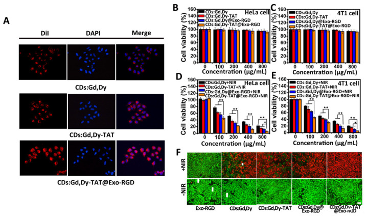
In vitro tumor cell targeting, nucleus penetration, cytotoxicity and photothermal performance. (A) DiI dye (red fluorescence)-labeled CDs:Gd,Dy, CDs:Gd,Dy-TAT or CDs:Gd,Dy-TAT@Exo-RGD was incubated with HeLa cells for 24 h, and DAPI was applied to dye the nucleus. The cell uptake was photographed through a fluorescence microscope, scale bar: 30 µm. (B–E) Viabilities of HeLa cells and 4T1 cells co-incubated with CDs:Gd,Dy, CDs:Gd,Dy-TAT, CDs:Gd,Dy@Exo-RGD or CDs:Gd,Dy-TAT@Exo-RGD of different concentrations (CDs:Gd,Dy: 0, 100, 200, 400 and 800 μg·mL−1) with/without exposure to laser (1.6 W·cm−2, 8 min). (F) Fluorescence images of Exo-RGD, CDs:Gd,Dy, CDs:Gd,Dy-TAT, CDs:Gd,Dy@Exo-RGD or CDs:Gd,Dy-TAT@Exo-RGD treated HeLa cells stained with Calcein AM and PI before and after laser irradiation (1.6 W·cm−2, 8 min), scale bar: 50 µm, * p < 0.05, ** p < 0.01.

