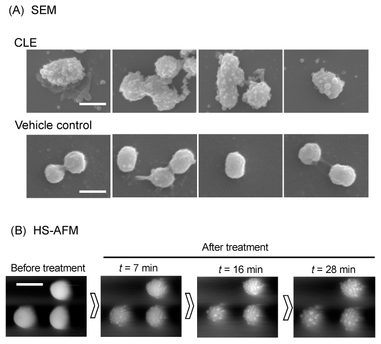
Figure 3.
Morphological change of P. gingivalis cells. (A) SEM analysis. P. gingivalis cells were treated with CLE at MIC or vehicle control for 30 min. Four different areas of CLE- and vehicle control-treated cells were shown. Scale: 1000 nm. (B) High-speed atomic force microscopy (HS-AFM) analysis. P. gingivalis cell morphology was monitored with a BIXAM system (Olympus) at nanometer scale. P. gingivalis cells were immobilized on glass slides and treated with CLE at 4-fold MIC (64 µg/mL). Nanometer-scale dynamics were continuously monitored. Shown are images of the cells before (Pre) and 7, 16, and 28 min after treatment with curry leaf (t = 7, 16, and 28). See also Supplemental Movies S1 and S2.

