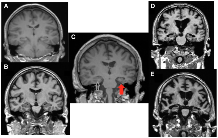
Figure 3.
Magnetic resonance imaging, coronal view. (A) Normal brain in a young subject; (B) Normal brain in an elderly subject; (C) Temporal brain atrophy in a 68-year-old patient with asymmetric severe hearing loss (red arrow). The white arrow shows the normal temporal lobe; (D) Diffused brain atrophy in a 72-year-old patient affected by cognitive decline; (E) Diffused brain atrophy in a 65-year-old patient with Alzheimer’s Disease [5].

