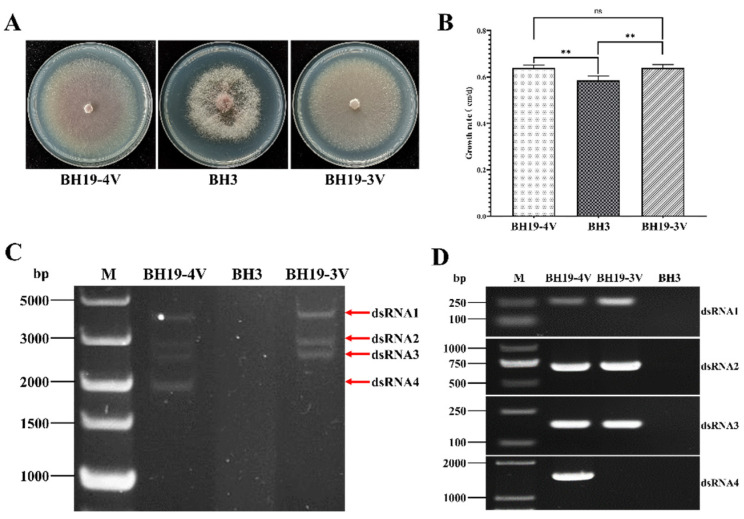
Figure 6.
(A) Colony morphology of F oxysporum strains BH3, BH19-3V and BH19-4V. (B) The growth rate of strains BH3, BH19-3V and BH19-4V. “**” represents significant difference (p < 0.05), “ns” represents no significant difference. (C) Agarose gel electrophoresis of the dsRNA-enriched extract obtained by cellulose column chromatography from strains BH3, BH19-3V and BH19-4V. (D) The detection of each segment of FoAV1 in strains BH3, BH19-3V and BH19-4V by RT-PCR amplification.

