Figure 2.
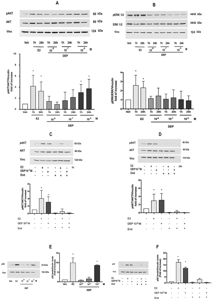
DEP activates nuclear and extra-nuclear rapid ERα signals. DEP dose-dependent effect on phosphorylation of AKT (A) and ERK1/2 (B) analyzed at short (1 h) and long (24 h) time exposures. Data show the Western blot image representative of at least four different experiments (upper panel) and the densitometric analysis (bottom panel). Western blot analysis of pAKT levels (upper panel) and densitometric analysis (bottom panel) in MCF-7 treated with DEP 10−5 M for 1 h (C) or 24 h (D) with or without cell pre-treatment with specific ERα inhibitor endoxifen (End; 10−6 M, 1 h before). Western blot (left panel) and corresponding densitometric analysis (right panel) of the E2-responsive protein pS2 in MCF-7 cells treated for 24 h with a dose curve of DEP (10−9, 10−7, 10−5 M) (E) or with DEP 10−5 M in the presence or absence of End (10−6 M, 1 h pre-treatment) (F). E2 in the presence and/or absence of End pre-treatment was used as internal positive control throughout all the experiments. The amount of protein was normalized in comparison with vinculin levels or with total AKT or total ERK1/2 and vinculin levels. Data are means ± SD of at least four experiments. p < 0.01 was determined by the ANOVA test followed by the Bonferroni post-test vs. Veh (-,-,-) (*).

