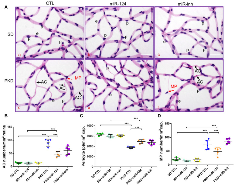
Figure 3.
miR-124 ameliorates pericyte loss and reduces vasoregression in PKD retinae. Retinal morphometry was measured in SD and PKD rats treated with or without miR-124. Two-month-old SD and PKD rats were treated with 25 pmol of control microRNA (CTL), an miR-124 mimic, or miR-124 inhibitor (miR-inh) for 4 weeks. (A) Representative images of PAS and hematoxylin-stained retinal vasculature from retinal digest preparation taken by an Olympus BX51 microscope. Black arrows indicate acellular capillaries (ACs), red arrows indicate migrating pericytes (MPs), p = pericyte, e = endothelial cell, and scale bars = 50 µm. (B–D) Quantification of acellular capillaries (number of ACs/mm2 retinal area) (B); quantification of pericytes (number of pericytes/mm2 retinal area) (C); and quantification of migrating pericytes (number of MP/mm2 retinal area) (D) were analyzed using CellF software from Olympus. (B–D) n = 5 and *** p < 0.001 (one-way ANOVA with Tukey’s multiple comparisons test).

