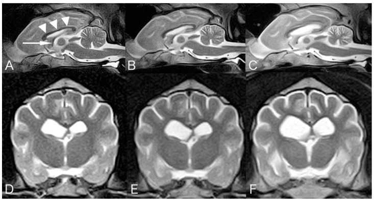
Figure 1.
Mid-sagittal T2-weighted magnetic resonance images at the age of 2 years and 11 months (A), 4 years and 7 months (B), and 5 years and 8 months (C). Transverse T2-weighted MR images at the level of the interventricular foramen at the time of 2 years and 11 months (D), 4 years and 7 months (E), and 5 years and 8 months (F). Progressive symmetrical atrophy was observed in the forebrain, cerebellum, brainstem, and cranial cervical spinal cord. Corpus callosum (arrowhead) and fornix (arrow) show gradual thinning.

