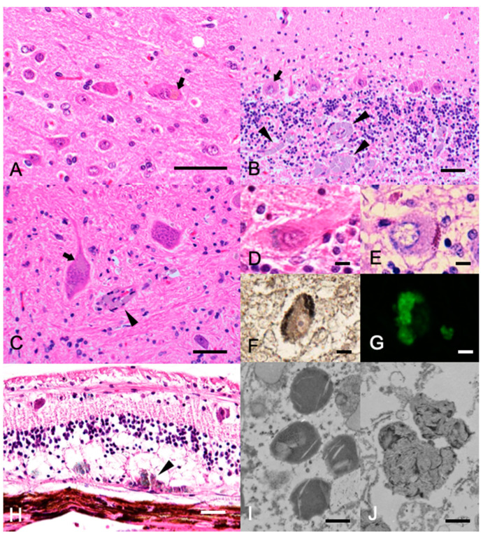
Figure 2.
Histopathology of the (A–C) Hematoxylin and eosin stain of the cerebrum (A), cerebellum (B), and neuronal nuclei of the pons (C). Bars = 50 µm. Light-brown pigments were frequently observed in the cytoplasm of large neurons (arrows). Note the pigment-phagocytosing macrophage infiltration in the perivascular region (arrowheads). (D–F) Histochemical staining of the cerebellum. Bars = 10 µm. The accumulated pigments in the Purkinje cells appeared blue in Luxol fast blue-hematoxylin and eosin (LFB-HE) stain (D), purple in periodic acid–Schiff (PAS) stain (E), and black in Sudan Black B stain (F). (G) Fluorescence microscopy image of the cerebellum. The accumulated pigments in the Purkinje cells showed green autofluorescence. (H) LFB-HE stain of the retina. Bar = 50 µm. The pigments were observed in the cytoplasm of the neuronal layer, and many macrophages phagocytosing the pigment were found at the border with the sclera (arrowheads). (I,J) Transmission electron microscopy of the storage bodies in neurons from the cerebellar Purkinje cells. Bar = 0.5 µm. The bodies showed membrane-like components.

