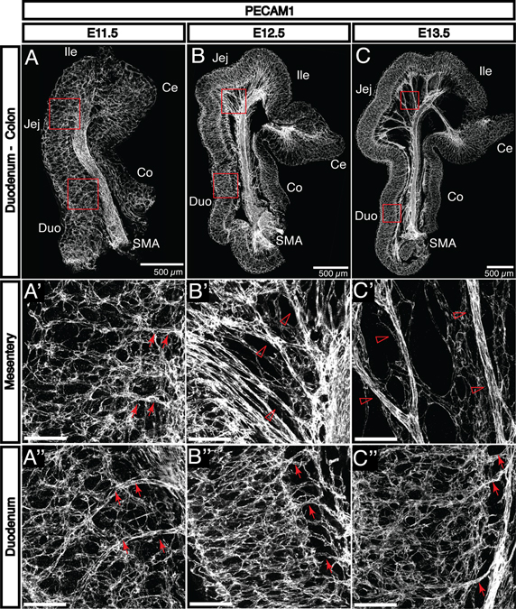
Fig. 2. The primary enteric capillary plexus is established at E11.5 and begins to remodel by E13.5.
A-C: PECAM1 staining reveals the development of embryonic vasculature in the small and large intestines from E11.5 to E13.5. A’-A”: The initial capillary plexus is uniform throughout the small intestine at E11.5. The mesentery is not yet detectable, and the superior mesenteric artery (SMA) is connected to the submucosal vascular plexus directly via capillaries (arrows). B’: As the gut tube lengthens at E12.5, it separates from the SMA to create the mesentery. The SMA is initially connected to the submucosal vasculature by smaller vessels (open arrowheads). C’: These vessels progressively remodel to form larger-diameter vessels which are observable by E13.5 (open arrowheads). B”: At E12.5 in the duodenum, the submucosal vasculature is connected to the mesenteric vasculature by small capillaries perpendicular to the length of the gut tube (arrows). C”: By E13.5, these vessels undergo a similar remodeling process to become recognizable as vasa recta (arrows). Notably, this process occurs first in the duodenum followed by the jejunum and the ileum. Duo, duodenum; Jej, jejunum; Ile, ileum; Ce, cecum; Co, colon. Scale bars = 100 μm except where indicated.

