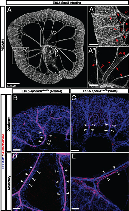
Fig. 3. Enteric arteries and veins adopt a stereotypical hierarchical pattern by E15.5.
A: By E15.5, the gut tube has completely separated from the SMA and the submucosal vasculature is connected to the SMA and SMV by both mesenteric vessels and vasa recta. A’: Vasa recta are readily apparent throughout the small intestine. These arteries (arrows) and veins (open arrowheads) are aligned and connect the submucosal vascular plexus with mesenteric vessels. A”: Mesenteric arteries (arrows) and veins (open arrowheads) are distinguishable by morphology. B-E: The expression of arterial (arrows) and venous (open arrowheads) molecular markers is visualized using LacZ reporter strains. Both types of vessels pattern adjacent to one another throughout intestinal development both in the mesentery and as they penetrate the gut wall. Scale bars = 100 μm except where indicated.

