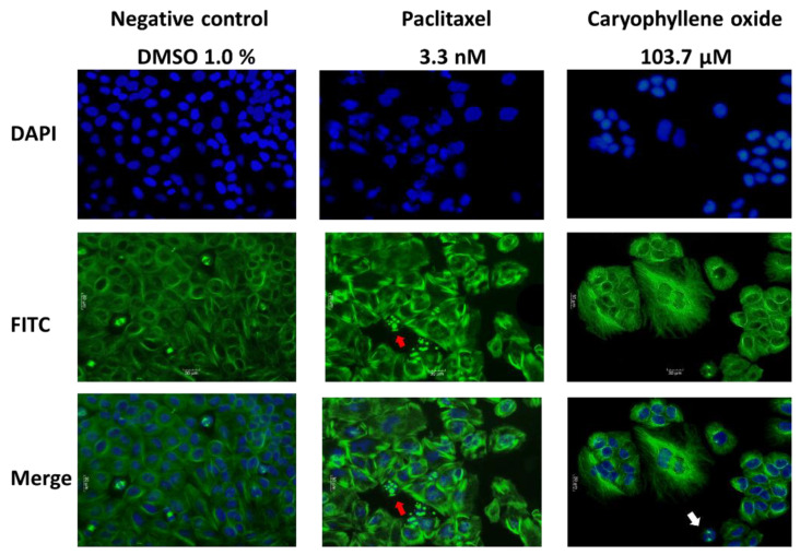
Figure 4.
Morphological changes in PC-3 after 24 h of OXC and Paclitaxel exposure. Immunofluorescence micrographs showing the nuclear (in blue) and microtubular (in green) effects in PC-3 cancer cells. Paclitaxel-treated cells displayed a moderate destabilization of the microtubules, an increase in cell size, the appearance of some apoptotic bodies (marked with a red arrow) that were not observed in the negative control cells treated with 1% DMSO, and the formation of abnormal nuclear morphologies. Otherwise, cells treated with OXC were visibly affected at the microtubules, considerably increasing the size of the cells, presenting cellular clusters, variable nuclear morphologies, and leading to the emergence of apoptotic cells (white arrow).

