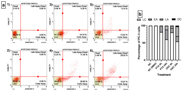
Figure 5.
Quantitative detection of Annexin V/7-AAD on PC-3 cells. (a) Dot plots of OXC and Paclitaxel treatments in the Annexin V/7-AAD assay. (1) Negative control, cells without any treatment. (2) Negative control, cells exposed to 1% DMSO. (3) Cells exposed to Paclitaxel for 8 h. (4) Cells exposed to Paclitaxel for 24 h. (5) Cells exposed to OXC for 8 h. (6) Cells exposed to OXC for 24 h. (b) Bar chart representing the distribution of the PC-3 cells detected by Annexin V/7-AAD following OXC and Paclitaxel exposure. Cells located in the lower right stained with Annexin V were defined as early apoptotic (EA), and Annexin V and 7-AAD double-stained cells were defined as late apoptotic (LA), located in the upper right. In the lower left are cells negative to both dyes, defined as live cells (LC), and cells in the upper left were stained only with 7-AAD, and correspond to non-apoptotic dead cells or nuclear debris.

