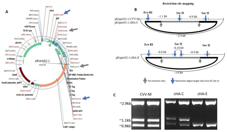
Figure 5.
RFLP verification of the HA cloned onto the pExpreS2-1 plasmid. (A) Plasmid map detailing HA insertion sites between NotI and XmaI (grey arrows or grey outline B) and existing EcoRI and SacII restriction sites (blue arrows). (B) Representation of EcoRI and SacII restriction digest sites showing anticipated electrophoretic band patterns. (C) Restriction digest confirming expected electrophoretic band patterns: SacII cuts twice the plasmid with the CVV-M and cHA-C at similar sites and EcoRI cuts once, hence the three similar band patterns; cHA-E on the other hand, displays 3 SacII restriction sites, in addition to one EcoRI site, leaving two major bands. Primers were also designed to target the flanks of the HA insertion point on the plasmid. These primers as well as the cloned pExpreS2-1 plasmids were outsourced for sequencing by GeneArt Gene Synthesis (Thermo Fisher Scientific, UK). Returned sequences confirmed the HA orientation on the plasmid (not shown).

