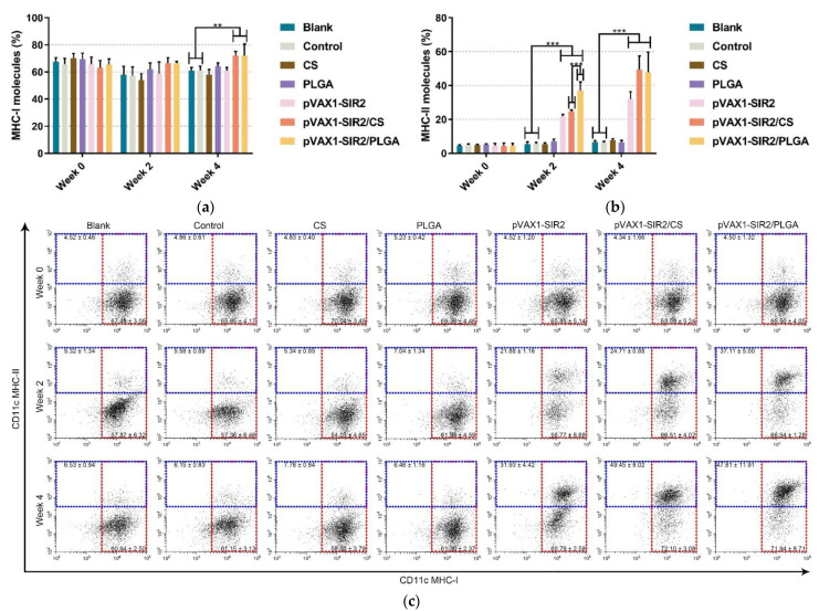
Figure 8.
Analysis of MHC molecules on the surface of DCs at week zero, two, and four. The expression of MHC-I and MHC-II molecules was evaluated by flow cytometry. Bar graph showed the ratio of MHC-I (a) and MHC-II (b) molecules on splenic DCs, and the dot plots (c) showed the percentage of CD11c+MHC-I+ and CD11c+MHC-II+ cells. Each sample was investigated once, and values were analyzed by one-way ANOVA analysis followed by Dunnett’s test. Values between the pVAX1-SIR2/CS and pVAX1-SIR2/PLGA group were estimated by the independent t-test. Values were shown as mean ± SD (n = 5). ** p < 0.01 and *** p < 0.001 compared with blank or control group.

