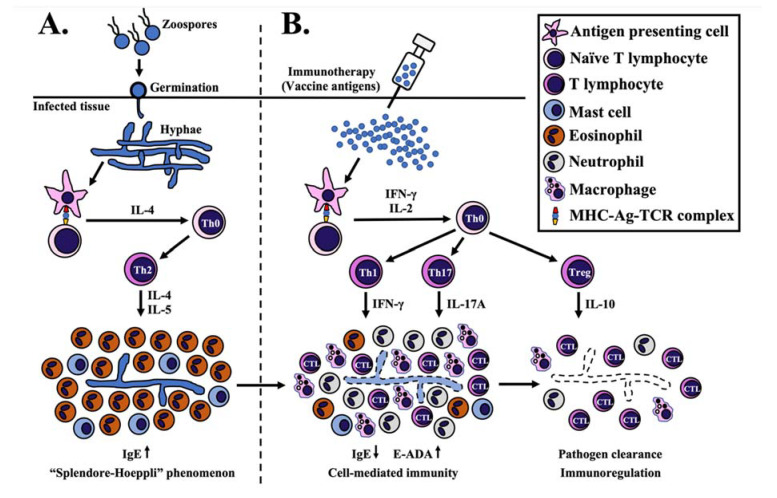
Figure 1.
Proposed mechanism of P. insidiosum antigen (PIA) immunotherapy. (A) P. insidiosum’s zoospores (asexual stage) attach and germinate as hyphae into the host tissue during natural infection. Antigen-presenting cells (APC) process and present the P. insidiosum antigens to naïve T lymphocytes via major histocompatibility complex-antigen-T cell receptor complex (MHC-Ag-TCR). This interaction elevates some cytokines (mainly IL-4) to differentiate and clonally proliferate a naïve CD4+ T (Th0) to T helper-2 (Th2) cell, which, in turn, produces IL-4 and IL-5 to attract and activate eosinophils and mast cells. The eosinophils surround the P. insidiosum, creating the histological phenomenon called “Splendore-Hoeppli”. (B) Processed antigens (prepared from the crude extract of P. insidiosum) lead to forming the MHC-Ag-TCR complex that induces the release of some cytokines, mainly IFN-ϒ and IL-2. IFN-ϒ promotes differentiation and clonal proliferation of a Th0 to T helper-1 (Th1) cell. Th1 cell-produced IFN-ϒ attracts macrophages and cytotoxic T lymphocytes (CTL) to the infection site for eliminating the pathogen. The MHC-Ag-TCR complex also facilitates the differentiation of a Th0 to T helper 17 (Th17) cell to produce IL-17A and accumulate more macrophages and neutrophils at the infection area. On the other hand, IL-2 promotes the differentiation of a Th0 to regulatory T (Treg) cell to regulate or suppress an excessive immune response through the release of IL-10. See the text for the details and the references to the proposed mechanism of PIA immunotherapy.

